Fetal Circulation
How does the fetal circulatory system work?
During pregnancy, the fetal circulatory system works differently than after birth:
-
The fetus is connected by the umbilical cord to the placenta. This is the organ that develops and implants in the mother's uterus during pregnancy.
-
Through the blood vessels in the umbilical cord, the fetus gets all needed nutrition and oxygen. The fetus gets life support from the mother through the placenta.
-
Waste products and carbon dioxide from the fetus are sent back through the umbilical cord and placenta to the mother's circulation to be removed.
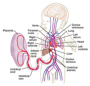
The fetal circulatory system uses three shunts. These are small passages that direct blood that needs to be oxygenated. The purpose of these shunts is to bypass the lungs and liver. That's because these organs will not work fully until after birth. The shunt that bypasses the lungs is called the foramen ovale. This shunt moves blood from the right atrium of the heart to the left atrium. The ductus arteriosus moves blood from the pulmonary artery to the aorta.
Oxygen and nutrients from the mother's blood are sent across the placenta to the fetus. The enriched blood flows through the umbilical cord to the liver and splits into three branches. The blood then reaches the inferior vena cava. This is a major vein connected to the heart. Most of this blood is sent through the ductus venosus. This is also a shunt that lets highly oxygenated blood bypass the liver to the inferior vena cava and then to the right atrium of the heart. A small amount of this blood goes straight to the liver to give it the oxygen and nutrients it needs.
Waste products from the fetal blood are transferred back across the placenta to the mother's blood.

Inside the fetal heart
-
Blood enters the right atrium. This is the chamber on the upper right side of the heart. When the blood enters the right atrium, most of it flows through the foramen ovale into the left atrium.
-
Blood then passes into the left ventricle. This is the lower chamber of the heart. Blood then passes to the aorta. This is the large artery coming from the heart.
-
From the aorta, blood is sent to the heart muscle itself and to the brain and arms. After circulating there, the blood returns to the right atrium of the heart through the superior vena cava. Very little of this less oxygenated blood mixes with the oxygenated blood. Instead of going back through the foramen ovale, it goes into the right ventricle.
-
This less oxygenated blood is pumped from the right ventricle into the pulmonary artery. A small amount of the blood continues on to the lungs. Most of this blood is shunted through the ductus arteriosus to the descending aorta. This blood then enters the umbilical arteries and flows into the placenta. In the placenta, carbon dioxide and waste products are released into the mother's circulatory system. Oxygen and nutrients from the mother's blood are released into the fetus's blood.
At birth, the umbilical cord is clamped, and the baby no longer gets oxygen and nutrients from the mother. With the first breaths of life, the lungs start to expand. As the lungs expand, the alveoli in the lungs are cleared of fluid. An increase in the baby's blood pressure and a major reduction in the pulmonary pressures reduce the need for the ductus arteriosus to shunt blood. These changes help the shunt close. These changes raise the pressure in the left atrium of the heart. They also lower the pressure in the right atrium. The shift in pressure stimulates the foramen ovale to close.
Blood circulation after birth
The closure of the ductus arteriosus, ductus venosus, and foramen ovale completes the change of fetal circulation to newborn circulation.
Online Medical Reviewer:
Heather M Trevino BSN RNC
Online Medical Reviewer:
Irina Burd MD PhD
Online Medical Reviewer:
Tennille Dozier RN BSN RDMS
Date Last Reviewed:
8/1/2023
© 2000-2026 The StayWell Company, LLC. All rights reserved. This information is not intended as a substitute for professional medical care. Always follow your healthcare professional's instructions.