Mammography
What is mammography (mammogram)?
Mammography is an x-ray examination of the breast. It is used to detect and diagnose breast disease in women who either have breast problems such as a lump, pain, or nipple discharge, as well as for women who have no breast complaints. The procedure allows detection of breast cancers, benign tumors, and cysts before they can be detected by palpation (touch).
Mammography cannot prove that an abnormal area is cancer, but if it raises a significant suspicion of cancer, tissue will be removed for a biopsy. Tissue may be removed by needle or open surgical biopsy and examined under a microscope to determine if it is cancer.
The development of digital mammography technology allows for improved breast imaging, in particular, for women less than 50 years of age, women with dense breast tissue, or women who are premenopausal or perimenopausal. Digital mammography provides electronic images of the breasts that can be enhanced by computer technology, stored on computers, and even transmitted electronically in situations where remote access to the mammogram is required. The procedure for a digital mammography is basically performed the same way as a standard mammogram.
With computer-aided detection (CAD) systems, a digitized mammographic image from a conventional film mammogram or a digitally acquired mammogram is analyzed for masses, calcifications, or areas of abnormal density that may indicate the presence of cancer. The images are highlighted by the CAD system for further analysis by the radiologist.
What is an x-ray?
X-rays use invisible electromagnetic energy beams to produce images of internal tissues, bones, and organs on film. Standard x-rays are performed for many reasons, including diagnosing tumors or bone injuries.

X-rays are made by using external radiation to produce images of the body, its organs, and other internal structures for diagnostic purposes. X-rays pass through body structures onto specially-treated plates (similar to camera film) and a "negative" type picture is made (the more solid a structure is, the whiter it appears on the film).
Anatomy of the breasts:
Each breast has 15 to 20 sections, called lobes, which are arranged like the petals of a daisy. Each lobe has many smaller lobules, which end in dozens of tiny bulbs that can produce milk.
The lobes, lobules, and bulbs are all linked by thin tubes called ducts. These ducts lead to the nipple in the center of a dark area of skin called the areola. Fat fills the spaces between lobules and ducts.
There are no muscles in the breast, but muscles lie under each breast and cover the ribs.
Each breast also contains blood vessels and vessels that carry lymph. The lymph vessels lead to small bean-shaped organs called lymph nodes, clusters of which are found under the arm, above the collarbone, and in the chest, as well as in many other parts of the body.

What are the different types of mammograms?
A screening mammogram is an x-ray of the breast used to detect breast changes in women who have no signs of breast cancer. It usually involves two x-rays of each breast. Using a mammogram, it is possible to detect a tumor that cannot be felt.
A diagnostic mammogram is an x-ray of the breast used to diagnose unusual breast changes, such as a lump, pain, nipple thickening or discharge, or a change in breast size or shape.
A diagnostic mammogram is also used to evaluate abnormalities detected on a screening mammogram. It is a basic medical tool and is appropriate in the work-up of breast changes, regardless of a woman's age.
Mammography has been used for about 30 years, and in the past 15 years technical advancements have greatly improved both the technique and results. Today, specialized equipment, used only for breast x-rays, produce studies that are high in quality but low in radiation dose.
Mammography may be used either for screening or to make a diagnosis. Women older than 25 years should undergo diagnostic mammography if they have symptoms such as a palpable lump, breast skin thickening or indentation, nipple discharge or retraction, erosive sore of the nipple, or breast pain.
A mammogram may be used to evaluate breast pain when physical examination and history are not conclusive. Women with breasts that are dense, "lumpy," and/or very large may be screened with mammography, as physical examination may be difficult to perform.
Women who are at high risk for breast cancer or with a history of breast cancer may be routinely screened with mammography.
Who should get a screening mammogram?
The following screening guidelines are for early detection of cancer in women who have no symptoms:
-
The American Cancer Society (ACS) recommends yearly screening for all women ages 40 and older. Women should talk with their doctors about their personal risk factors before making a decision about when to start getting mammograms or how often they should get them.
-
The ACS recommends clinical breast exams (CBEs) at least every three years for all women in their 20s and 30s. The ACS recommends annual CBEs for women ages 40 and older. Women should talk with their doctors about their personal risk factors and make a decision about whether they should have a CBE.
-
The ACS says BSEs are an option for women 20 and older as a means of familiarizing themselves with their breasts so they can notice changes more easily. Talking with your doctor about the benefits and limitations can help you decide if you should start performing BSEs.
-
Women who are at an increased risk (family history, genetic tendency, past breast cancer) should talk with their physicians about the benefits and limitations of starting mammography screening earlier, having additional tests (breast ultrasound, MRI), or having more frequent exams.
In addition, the following guidelines by age are recommended:
-
National Cancer Institute Guideline for Screening Mammography: Women in their 40s and older should have a screening mammogram on a regular basis, every one to two years.
-
American Cancer Society Guideline for Screening Mammography: Women 40 years of age and older should have a screening mammogram every year.
Consult your physician regarding the screening guidelines that are appropriate for you.
What conditions does a mammogram show?

A mammogram helps to identify the following conditions:
-
calcifications - tiny mineral deposits within the breast tissue. There are two categories of calcifications:
-
macrocalcifications - coarse calcium deposits that usually indicate degenerative changes in the breasts, such as the following:
-
microcalcifications - tiny (less than 1/50 of an inch) specks of calcium. When many microcalcifications are seen in one area, they are referred to as a cluster.
-
masses - may occur with or without associated calcifications, and may be due to different causes, including the following:
-
cyst - a non-cancerous collection of fluid in the breast. Cysts cannot be diagnosed by physical examination alone or by mammography alone. Either breast ultrasound or aspiration with a needle is required. If a mass is not a cyst, then further imaging may be necessary.
-
benign breast conditions - masses can be monitored with periodic mammography, but others may require immediate or delayed biopsy. About 80 percent of all breast changes that are biopsied are found to be benign (non-cancerous) when looked at under the microscope.
-
breast cancer
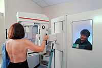
How is a mammogram performed?
Usually, a mammogram is done on an outpatient basis, although it can be part of inpatient care. There is no specific preparation for the examination. However, a woman should not wear deodorant, powders, or lotions under the arms on the day of the examination, as these substances can interfere with the images. If you have breast implants, be sure to tell your mammography facility that you have them when you make your appointment. You will need an x-ray technologist who is trained in working with patients with implants. This is important because breast implants can hide some breast tissue, which could make it difficult for the radiologist to see breast cancer when looking at your mammogram images.
Although each facility may have specific protocols in place, generally, a mammogram procedure follows this process:
-
The patient should describe any symptoms or problems to the technologist prior to the examination (if any).
-
The patient will undress from the waist up and will be given a gown to wear.
-
The patient will be positioned at the mammography unit, seated, standing, or lying down.
-
The breast will be positioned between two plates of the mammography unit, and pressure applied to compress the tissue. (This may produce temporary discomfort.) Breast compression is necessary in order to obtain the best image with the least amount of radiation possible.
-
The patient will be asked to hold her breath for a few seconds while the x-rays are taken.
-
The technologist will step behind a protective window and the image will be taken.
-
Each breast may be x-rayed at least two times from above and from the side positions to produce the films for the physician to review.
-
After the x-rays are made, the patient will be asked to wait for a short time until the radiologist can review the films to determine if additional x-rays are necessary.
-
The examination process takes approximately 20 to 30 minutes.