What is a biopsy?
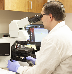
A biopsy is a procedure to remove a tissue sample or cells from the body. The sample is then checked under a microscope. Some biopsies can be done in a healthcare provider's office. Others need to be done in a hospital. In addition, some biopsies can be done with an anesthetic to numb just the local area. Others may need sedation or even full anesthesia that puts you completely asleep during the procedure.
Biopsies are often done to find out if a tumor is cancer. They are also done to find the cause of an unexplained sore, mole, infection, or inflammation.

Types of biopsies
Endoscopic biopsy
This type of biopsy is done through a fiber-optic endoscope. This is a long, thin tube that has a close-focusing telescope on the end for viewing. The scope may be passed through a natural body opening (for example, rectum or mouth). Or it may be passed through a small cut (incision). It's used to look at the organ in question for abnormal or suspicious areas so that a small amount of tissue can be removed for study. Endoscopic procedures are named for the organ or body area to be viewed or treated. The healthcare provider can put the endoscope into the:
-
Gastrointestinal tract (alimentary tract endoscopy)
-
Bladder (cystoscopy)
-
Belly or abdominal cavity (laparoscopy)
-
Joint cavity (arthroscopy)
-
Middle part of the chest (mediastinoscopy)
-
Trachea and bronchial system (laryngoscopy and bronchoscopy)
Bone marrow biopsy
Bone marrow aspiration or biopsy means taking a small amount of bone marrow fluid (aspiration) or solid bone marrow tissue (called a core biopsy). The tissue is often taken from the back of the hipbones. It's then checked for the number, size, and maturity of blood cells or abnormal cells.
Excisional or incisional biopsy
A surgical biopsy is often used when a wider or deeper part of the tissue is needed. Using a surgical knife (scalpel), a full thickness of skin or all or part of a large tumor may be removed. The wound is sewn closed with surgical thread.
When the entire tumor is removed, it's called an excisional biopsy. If only part of the tumor is removed, it's called an incisional biopsy. For instance, excisional biopsy is the method often preferred when melanoma is suspected. Both types of biopsies can be done by using local or regional anesthesia. If the tumor is inside the chest or belly, general anesthesia is used. In some cases, surgeons will take an excisional or incisional biopsy that goes right to the pathologist while the person is still under anesthesia. This is to make sure a tumor is fully removed. One example of this is during Mohs surgery that is commonly performed on the face.
Fine needle aspiration (FNA) biopsy
This type of biopsy uses a thin needle and syringe to remove very small pieces from a tumor. Local anesthetic is sometimes used to numb the area. The test rarely causes much discomfort. It leaves no scar.
FNA is not used for diagnosis of a suspicious mole. It may be used to biopsy large lymph nodes near a melanoma to see if the melanoma has spread. Some breast and thyroid tumors may be studied through FNA. A CT scan may be used to guide a needle into a tumor in an internal organ, such as the lung or liver. An ultrasound, fluoroscopy (a type of continuous X-ray), or other study may also be used to help guide the needle. A core biopsy is like an FNA. But it uses a larger needle for a larger tissue sample.
Punch biopsy
Punch biopsies take a deeper sample of skin. They use a biopsy tool that removes a short cylinder, or "apple core," of tissue. After a local anesthetic, the tool is turned on top of the skin until it cuts through all the skin layers.
Shave biopsy
This type of biopsy removes the top layers of skin by shaving it off. Shave biopsies are used to diagnose some basal cell or squamous cell skin cancers as well as other skin sores. But they are not advised for suspected skin melanomas. Shave biopsies are also done with a local anesthetic.
Reflectance confocal microscopy (RCM)
This method lets the healthcare provider look at an abnormal area of skin to a certain depth. But it doesn't cut into the skin or remove a skin sample. RCM is used widely in Europe. It's now available in some centers in the U.S.