Mammogram Procedure
(Mammography, Breast X-ray)
Procedure Overview
What is a mammogram?
A mammogram is an x-ray examination of the breast. It is used to detect and diagnose breast disease in women who either have breast problems such as a lump, pain, or nipple discharge, as well as for women who have no breast complaints. The procedure allows detection of breast cancers, benign tumors, and cysts before they can be detected by palpation (touch).
Mammography cannot prove that an abnormal area is cancer, but if it raises a significant suspicion of cancer, tissue will be removed for a biopsy. Tissue may be removed by needle or open surgical biopsy and examined under a microscope to determine if it is cancer.
Mammography has been used for about 30 years, and in the past 15 years technical advancements have greatly improved both the technique and results. Today, dedicated equipment, used only for breast x-rays, produces studies that are high in quality but low in radiation dose. Radiation risks are considered to be negligible.

The development of digital mammography technology allows for improved breast imaging, in particular, for women less than 50 years of age, women with dense breast tissue, and women who are premenopausal or perimenopausal. Digital mammography provides electronic images of the breasts that can be enhanced by computer technology, stored on computers, and even transmitted electronically in situations where remote access to the mammogram is required. The procedure for a digital mammography is basically performed the same way as a standard mammogram.
With computer-aided detection (CAD) systems, a digitized mammographic image from a conventional film mammogram or a digitally acquired mammogram is analyzed for masses, calcifications, or areas of abnormal density that may indicate the presence of cancer. The images are highlighted by the CAD system for further analysis by the radiologist.
What are the different types of mammograms?
According to the National Cancer Institute:
-
Screening mammogram
A screening mammogram is an x-ray of the breast used to detect breast changes in women who have no signs or symptoms of breast cancer. It usually involves two x-rays of each breast. Using a mammogram, it is possible to detect a tumor that cannot be felt.
-
Diagnostic mammogram
A diagnostic mammogram is an x-ray of the breast used to diagnose unusual breast changes, such as a lump, pain, nipple thickening or discharge, or a change in breast size or shape.
A diagnostic mammogram is also used to evaluate abnormalities detected on a screening mammogram. It is a basic medical tool and is appropriate in the workup of breast changes, regardless of a woman's age.
What is an x-ray?
X-rays use invisible electromagnetic energy beams to produce images of internal tissues, bones, and organs on film. Standard x-rays are performed for many reasons, including diagnosing tumors or bone injuries.

X-rays are made by using external radiation to produce images of the body, its organs, and other internal structures for diagnostic purposes. X-rays pass through body structures onto specially-treated plates (similar to camera film) and a "negative" type picture is made (the more solid a structure is, the whiter it appears on the film).
Anatomy of the Breasts
Each breast has 15 to 20 sections, called lobes, which are arranged like the petals of a daisy. Each lobe has many smaller lobules, which end in dozens of tiny bulbs that can produce milk.
The lobes, lobules, and bulbs are all linked by thin tubes called ducts. These ducts lead to the nipple in the center of a dark area of skin called the areola. Fat fills the spaces between lobules and ducts.
There are no muscles in the breast, but muscles lie under each breast and cover the ribs.
Each breast also contains blood vessels and vessels that carry lymph. The lymph vessels lead to small bean-shaped organs called lymph nodes, clusters of which are found under the arm, above the collarbone, and in the chest, as well as in many other parts of the body.
Reasons for the Procedure
Mammography may be used either for screening or to make a diagnosis. Women older than 25 years should undergo diagnostic mammography if they have symptoms such as a palpable lump, breast skin thickening or indentation, nipple discharge or retraction, erosive sore of the nipple, or breast pain.
A mammogram may be used to evaluate breast pain when physical examination and history are not conclusive. Women with breasts that are dense, "lumpy," and/or very large may be screened with mammography, as physical examination may be difficult to perform.
Women who are at high risk for breast cancer or with a history of breast cancer may be routinely screened with mammography.
There may be other reasons for your physician to recommend a mammography.
Who should get a screening mammogram?
The following screening guidelines are for early detection of cancer in women who have no symptoms:
-
The American Cancer Society (ACS) recommends yearly screening for all women ages 40 and older. Women should talk with their doctors about their personal risk factors before making a decision about when to start getting mammograms or how often they should get them.
-
The ACS recommends clinical breast exams (CBEs) at least every three years for all women in their 20s and 30s. The ACS recommends annual CBEs for women ages 40 and older. Women should talk with their doctors about their personal risk factors and make a decision about whether they should have a CBE.
-
The ACS says BSEs are an option for women 20 and older as a means of familiarizing themselves with their breasts so they can notice changes more easily. Talking with your doctor about the benefits and limitations can help you decide if you should start performing BSEs.
-
Women who are at an increased risk (family history, genetic tendency, past breast cancer) should talk with their physicians about the benefits and limitations of starting mammography screening earlier, having additional tests (breast ultrasound, MRI), or having more frequent exams.
In addition, the following guidelines by age are recommended:
-
National Cancer Institute Guideline for Screening Mammography:
Women in their 40s and older should have a screening mammogram on a regular basis, every one to two years.
-
American Cancer Society Guideline for Screening Mammography:
Women 40 years of age and older should have a screening mammogram every year.
Consult your physician regarding the screening guidelines that are appropriate for you.
Risks of the Procedure
You may want to ask your physician about the amount of radiation used during the procedure and the risks related to your particular situation. It is a good idea to keep a record of your past history of radiation exposure, such as previous scans and other types of x-rays, so that you can inform your physician. Risks associated with radiation exposure may be related to the cumulative number of x-ray examinations and/or treatments over a long period of time.
If you are pregnant or suspect that you may be pregnant, you should notify your physician. Radiation exposure during pregnancy may lead to birth defects. If it is necessary for you to have a mammogram, special precautions will be made to minimize the radiation exposure to the fetus.
Mammograms may be more difficult to interpret in women younger than 30 years of age, due to the increased density of their breast tissue.
Some discomfort may be felt as the breast is compressed against the x-ray plate during the procedure. This compression will not harm the breast, however.
There may be other risks depending upon your specific medical condition. Be sure to discuss any concerns with your physician prior to the procedure.
Certain factors or conditions may interfere with a mammogram. These include, but are not limited to, the following:
-
Talcum powder, deodorant, creams, or lotions applied under the arms or on the breasts
-
Breast implants, as they may prevent complete visualization of the breast. If you have breast implants, be sure to tell your mammography facility that you have them when you make your appointment. You will need an x-ray technologist who is trained in working with patients with implants. This is important because breast implants can hide some breast tissue, which could make it difficult for the radiologist to see breast cancer when looking at your mammogram images.
-
Previous breast surgery
-
Hormonal breast changes
Before the Procedure
-
Your physician will explain the procedure to you and offer you the opportunity to ask any questions that you might have about the procedure.
-
You may be asked to sign a consent form that gives permission to do the procedure. Read the form carefully and ask questions if something is not clear.
-
No fasting or sedation is required before the procedure.
-
If you are pregnant or suspect that you may be pregnant, you should notify your physician.
-
Notify your physician of all medications (prescribed and over-the-counter) and herbal supplements that you are taking.
-
Notify your physician if you have breast implants or if you are breastfeeding.
-
Dress in clothes that permit access to the area to be tested or that are easily removed.
-
Comparison with old mammograms is very important. If you are having a mammogram performed at a new facility, you may be asked to retrieve your previous mammograms from the previous facility.
-
Avoid using deodorant, perfume, powders, or ointment on the breast or underarm area on the day of the mammogram. It may interfere with the reading.
-
Patients with painful breasts may be asked to refrain from caffeinated food and beverages for five to seven days before testing.
-
Breasts are often tender the week before and during menstruation, so try to schedule your mammogram for one to two weeks after your period starts.
-
Based upon your medical condition, your physician may request other specific preparation.
During the Procedure
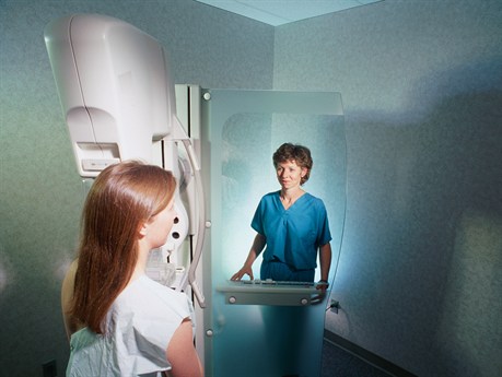
Mammogram
A mammogram may be performed on an outpatient basis or as part of your stay in a hospital. Procedures may vary depending on your condition and your physician's practices.
Generally, a mammogram follows this process:
-
You will be asked to remove any clothing, jewelry, or other objects that might interfere with the procedure.
-
You will be asked to remove clothing from your waist up, and will be given a gown to wear.
-
The technologist will ask you if you have noticed any lumps or other changes in either breast. If so, an adhesive marker will be placed on the spot(s) prior to the procedure.
-
You will stand in front of a mammography machine and one breast will be placed on the x-ray plate. In order to position the breast for optimal imaging, the technologist may examine and/or palpate the breast before placing it on the plate. An adhesive marker may be applied to any moles, scars, or other spots that might interfere with the breast image.
-
A separate flat plate, often made of plastic, will be brought down on top of the breast to compress it gently against the x-ray plate. Compression of the breast is required in order to minimize the amount of radiation used and to ensure optimal visualization of the breast tissue. You may feel some discomfort during this time.
-
You will be asked to hold your breath while the image is being taken.
-
The radiologic technologist will step behind a protective window while the image is taken.
-
Two pictures at different angles will be taken of each breast, requiring the breasts to be repositioned between pictures.
-
After the x-rays have been taken, you will be asked to wait while the films are examined by the radiologist to ensure that the films are clear and that no additional films are needed. If there is a question about any of the films, you may be asked to have additional films taken.
-
The examination process takes approximately 20 to 30 minutes.
While the mammogram itself causes no pain, the manipulation and compression of the breast being examined may cause some discomfort or pain, particularly in the case of a recent injury or invasive procedure such as surgery. The radiologic technologist will use all possible comfort measures and complete the procedure as quickly as possible to minimize any discomfort or pain.
After the Procedure
Generally, there is no special type of care following a mammogram. Your physician may give you additional or alternate instructions after the procedure, depending on your particular situation.
Online Resources
The content provided here is for informational purposes only, and was not designed to diagnose or treat a health problem or disease, or replace the professional medical advice you receive from your physician. Please consult your physician with any questions or concerns you may have regarding your condition.
This page contains links to other Web sites with information about this procedure and related health conditions. We hope you find these sites helpful, but please remember we do not control or endorse the information presented on these Web sites, nor do these sites endorse the information contained here.
American Cancer Society
American College of Radiology
American Society of Clinical Oncology
National Cancer Institute (NCI)
National Institutes of Health (NIH)
National Library of Medicine